
![]()
![]()
AFM: ATOMIC FORCE MICROSCOPE
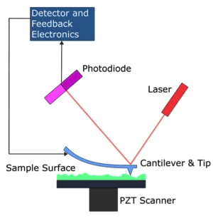 This
is a scanning microscope
and it
makes a scan of the material surface using a tip of atomic
dimensions, placed at the end of a tiny flexible rod. The movements
of the rod are induced by forces
of
inter-atomic interaction
at short-range
between
the tip of the microscope and the atoms of the sample surface; these
movements reveal information about the structure of the sample.
Basically, the tip of the rod is positioned on a flexible arm, called
cantilever,
at a distance from the sample surface lower than one nanometer, when
the sample is translated along the x-y plane below the tip, repulsion
forces or attractive forces are manifested between the sample surface
and the tip, with a consequent deflection of the tip in z direction.
The morphology of the sample surface is rebuilt by measuring the
deflection entity of the tip. In addition, the limitation to analyze
only conductive samples doesn't exist with this microscope, in fact a
wide variety of samples can be analyzed, for example: plastic, glass
and biomolecules.
This
is a scanning microscope
and it
makes a scan of the material surface using a tip of atomic
dimensions, placed at the end of a tiny flexible rod. The movements
of the rod are induced by forces
of
inter-atomic interaction
at short-range
between
the tip of the microscope and the atoms of the sample surface; these
movements reveal information about the structure of the sample.
Basically, the tip of the rod is positioned on a flexible arm, called
cantilever,
at a distance from the sample surface lower than one nanometer, when
the sample is translated along the x-y plane below the tip, repulsion
forces or attractive forces are manifested between the sample surface
and the tip, with a consequent deflection of the tip in z direction.
The morphology of the sample surface is rebuilt by measuring the
deflection entity of the tip. In addition, the limitation to analyze
only conductive samples doesn't exist with this microscope, in fact a
wide variety of samples can be analyzed, for example: plastic, glass
and biomolecules.
For
example, this spectacular image
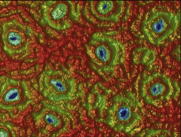 represents the bottom surface of a plant leaf, observed with an AFM.
The openings that are noticed on lower epidermis of the leaf are
called stomata;
they allow
gaseous exchange between the air and the interior of the leaf.
represents the bottom surface of a plant leaf, observed with an AFM.
The openings that are noticed on lower epidermis of the leaf are
called stomata;
they allow
gaseous exchange between the air and the interior of the leaf.
The
following figures give the microscope images of
the graphite, starting from a vision obtained with an optical
microscope
 (that uses visible light to
create a magnified image of an object), up to an image obtained with
recent scanning microscopes:
(that uses visible light to
create a magnified image of an object), up to an image obtained with
recent scanning microscopes:
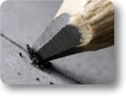 naked-eye
view;
naked-eye
view;
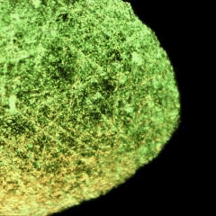 vision by an optical
microscope;
vision by an optical
microscope;
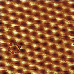 vision by a scanning AFM.